Glaucoma Treatment & Diagnosis — Advanced Eye Treatment at a Trusted, NABH-Accredited Eye Care Hospital
Expert glaucoma diagnosis, monitoring, laser treatment, and surgery — protecting the optic nerve and preserving your vision for life. Experienced glaucoma eye specialist doctors across 5 centres.
Glaucoma is called the silent thief of sight for one devastating reason — it destroys vision gradually and painlessly, with no warning until significant, irreversible damage has already occurred. At MediVision Eye Care Centre, our NABH-accredited eye care hospital offers comprehensive glaucoma diagnosis, advanced eye treatment, and lifelong monitoring to detect this condition early and protect your vision from further loss.
What Is Glaucoma — Why Is It Called the Silent Thief of Sight?
No pain, no redness, no obvious change in vision — until the damage is already permanent.
Glaucoma is a group of eye conditions in which the optic nerve — the vital neural cable that transmits visual signals from the eye to the brain — is progressively damaged. In most cases, this damage is driven by elevated intraocular pressure (IOP). Left undetected and untreated, glaucoma causes irreversible vision loss, beginning with peripheral vision and advancing to complete blindness.
What makes glaucoma so dangerous is its silence. In the most common form — primary open-angle glaucoma — there is no pain, no redness, and no obvious change in vision until the disease is advanced. Patients lose peripheral vision so gradually that the brain compensates, and they do not notice the loss until only central tunnel vision remains. By that point, the damage is permanent.
According to the World Health Organization, glaucoma is the second leading cause of irreversible blindness worldwide, responsible for approximately 8% of global blindness. Yet with timely diagnosis during a routine comprehensive eye checkup and appropriate advanced eye treatment, vision loss from glaucoma is largely preventable.
What Causes Glaucoma?
The eye continuously produces a clear fluid called aqueous humour, which nourishes and maintains the shape of the eye. This fluid flows through the anterior chamber and drains out through a specialised canal at the angle of the eye. When drainage is impaired, fluid accumulates and intraocular pressure rises — exerting mechanical pressure on the optic disc and progressively damaging nerve fibres. This process is typically painless and invisible until well advanced.
Normal tension glaucoma — where optic nerve damage occurs despite statistically normal IOP — is a well-recognised variant, which is why a comprehensive eye checkup for glaucoma involves optic nerve assessment alongside pressure measurement.
What Are the Different Types of Glaucoma — Which Type Do You Have?
Accurate type classification determines the most effective advanced eye treatment approach.
Primary Open-Angle Glaucoma (POAG)
The most common form worldwide. The drainage angle appears open and normal, but microscopic resistance within the trabecular meshwork impairs outflow, causing IOP to rise gradually. Symptoms are absent until significant peripheral vision loss — managed with drops, laser, and surgery.
Angle-Closure Glaucoma
The drainage canal is physically blocked by the iris. Acute angle-closure is a medical emergency — sudden severe eye pain, headache, nausea, blurred vision, and coloured haloes — requiring immediate treatment to prevent vision loss within hours.
Developmental Glaucoma
Occurs when the eye's drainage structures do not develop normally before or after birth. Congenital glaucoma presents from birth with enlarged, cloudy eyes and excessive tearing. Juvenile glaucoma affects children and young adults. Both typically require early surgical intervention.
Secondary Glaucoma
Develops as a consequence of another condition — steroid-induced (from prolonged eye drop, nasal spray, or oral steroid use), neovascular (secondary to diabetic retinopathy), traumatic/angle recession (following eye injury), or uveitic (from intraocular inflammation).
Who Is at Risk of Glaucoma — Do You Need a Glaucoma Eye Checkup?
Glaucoma has no symptoms in its early stages. Knowing your risk factors is your most powerful protection.
Our eye specialist doctors assess the following risk factors as part of every comprehensive eye checkup. The American Academy of Ophthalmology recommends that adults with multiple risk factors begin glaucoma screening before the age of 40.
Age
Risk increases significantly with age. Adults over 40 should have annual glaucoma screening — even without symptoms. After 60, risk escalates further.
Family History
A first-degree relative with glaucoma increases personal risk four to nine times — one of the strongest independent risk factors.
Refractive Errors
Both high myopia and high hyperopia are associated with increased glaucoma risk. High myopes should be screened at every visit.
Diabetes & Hypertension
Uncontrolled diabetes and high blood pressure can cause neovascular glaucoma, one of the most aggressive secondary forms.
Prolonged Steroid Use
In any form — drops, nasal sprays, ointments, or tablets — prolonged use without IOP monitoring is a significant preventable cause.
Vascular Factors
Conditions affecting blood flow to the optic nerve — migraine, Raynaud's phenomenon, obstructive sleep apnoea — are associated with normal tension glaucoma.
Prior Eye Trauma
Previous injury can damage the drainage angle, leading to angle recession glaucoma years later.
If any of these risk factors apply to you, book a glaucoma eye checkup at our eye care hospital today.
Book Your Eye CheckupHow Is Glaucoma Diagnosed at Our Eye Care Hospital?
Accurate diagnosis requires more than a single pressure test — it requires a complete structural and functional assessment.
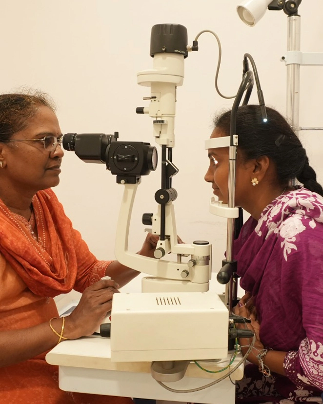
Since glaucoma causes no symptoms in its early stages, detection is almost entirely dependent on systematic screening during a comprehensive eye checkup. Our eye specialist doctors do not rely on a single test — glaucoma diagnosis requires correlation of multiple clinical findings to distinguish true disease from glaucoma suspects who require monitoring.
Intraocular Pressure (IOP) Measurement
Multiple tonometry methods — Non-Contact Tonometer for routine screening, Goldmann Applanation Tonometer as the gold-standard reference, and Perkins Tonometer for patients who cannot be positioned at the slit lamp.
Gonioscopy — Drainage Angle Assessment
Using a specialised contact lens, our glaucoma specialists directly visualise the drainage angle to classify open-angle versus angle-closure glaucoma and plan the correct treatment approach.
Optic Disc Examination & Photography
The optic nerve head is examined under dilated pupil — assessing cup-to-disc ratio, rim tissue, and structural changes. Colour and red-free photography documents baseline and progression.
OCT — Optic Disc & RNFL Analysis
Objective, quantitative measurement of the retinal nerve fibre layer and optic nerve head — detecting structural damage often before visual field changes appear.
Visual Field Testing — Humphrey Perimetry
The gold-standard instrument for mapping glaucoma-related visual field loss — quantifying the degree and pattern of peripheral vision damage and monitoring progression.
Pachymetry — Central Corneal Thickness
Corneal thickness affects IOP readings and is an independent risk factor for progression. Thin corneas are linked to underestimated IOP and greater susceptibility to damage.
What Are the Glaucoma Treatment Options at MediVision Eye Care Hospital?
Glaucoma cannot be cured — but with the right treatment and lifelong monitoring, vision loss can be halted.
Our eye specialist doctors develop personalised glaucoma treatment plans based on type, severity, rate of progression, patient age, and systemic health — following American Academy of Ophthalmology guidelines. Treatment is reviewed and adjusted at every follow-up, as glaucoma management is a lifelong commitment.
Medical Management — Eye Drops
- Prostaglandin analogues — first-line, once-daily, high-efficacy agents increasing uveoscleral outflow
- Beta-blockers — reduce aqueous production, used alone or in combination
- Alpha-agonists — reduce production and increase outflow
- Carbonic anhydrase inhibitors — drops or oral tablets for acute IOP lowering
- Combination drops — fixed formulations that improve regimen compliance
Eye drop compliance is the single most important determinant of treatment success. Skipping drops allows IOP to fluctuate and optic nerve damage to progress unimpeded.
Laser Treatment
Laser Peripheral Iridotomy (LPI) is the primary treatment for angle-closure glaucoma. Using an Nd:YAG laser, a small opening is created in the peripheral iris, providing an alternative drainage pathway and relieving the pupillary block — also performed prophylactically in narrow-angle eyes at risk.
Surgical Glaucoma Treatment
For patients in whom IOP remains uncontrolled despite maximum medical therapy and laser, surgical treatment is indicated:
Trabeculectomy
The gold-standard glaucoma surgery — creates a controlled filtering bleb through which aqueous drains, bypassing the blocked canal. Sustainably lowers IOP in most patients under local anaesthesia.
Glaucoma Drainage Device (AGV)
Ahmed Glaucoma Valve implantation places a small silicone tube draining aqueous to an external plate — used for complex cases including neovascular and uveitic glaucoma.
MIGS
Minimally Invasive Glaucoma Surgery — iStent, Hydrus, KDB and BANG goniotomy — addresses outflow resistance with minimal surgical trauma and rapid recovery, often combined with cataract surgery.
Combined Cataract & Glaucoma Surgery
For patients with both visually significant cataract and glaucoma requiring surgery — addressing both conditions in a single procedure, reducing overall surgical burden.
Cyclodestructive Procedures
For refractory glaucoma unresponsive to other treatments — cyclocryopexy and transscleral cyclo-photocoagulation reduce IOP by targeting the ciliary body.
Why Does Early Glaucoma Detection Change Everything?
Glaucoma damage is irreversible. Early detection is the only way to protect the vision you still have.
The optic nerve fibres destroyed by glaucoma do not regenerate. Every degree of visual field lost to glaucoma is permanent. This biological reality makes early detection — through regular comprehensive eye checkups — the single most powerful intervention available against glaucoma-related blindness.
What Does Early Glaucoma Diagnosis Achieve?
Preserves Remaining Vision
Treatment initiated early preserves significantly more visual function than treatment started after moderate or severe damage — often the difference between lifelong functional vision and severe impairment.
Simpler, Less Invasive Treatment
Early glaucoma responds well to drops or laser. Late-stage disease frequently requires surgery with higher risk and longer recovery.
Protects Quality of Life
Peripheral vision loss affects driving, mobility, fall risk, and independence — particularly in older patients. Timely treatment preserves what central vision alone cannot maintain.
Identifies Systemic Conditions
Glaucoma secondary to diabetes, hypertension, or steroid use prompts investigation of the underlying systemic condition — sometimes the first indication of uncontrolled disease.
Screens Family Members
A confirmed diagnosis in one family member is a strong indication for first-degree relatives to attend a glaucoma eye checkup.
Recommended Glaucoma Screening Schedule
| Patient Group | Recommended Frequency |
|---|---|
| Adults under 40, no risk factors | Every 2 years |
| Adults over 40 | Annually |
| Glaucoma suspect (elevated IOP / suspicious disc) | Every 6 months |
| Confirmed glaucoma — stable | Every 3–6 months |
| Confirmed glaucoma — under treatment adjustment | Every 1–3 months |
| Family history of glaucoma | Annually from age 35 |
| Steroid users (any route) | Every 3–6 months |
| High myopes (above -6.00D) | Annually |
Source: American Academy of Ophthalmology preferred practice pattern for glaucoma.
What Advanced Eye Treatment Technology Does MediVision Use for Glaucoma?
Accurate diagnosis and effective treatment planning depend on the right technology — and the experience to interpret it.
Humphrey Visual Field Analyser
The internationally recognised gold-standard for visual field assessment — baseline diagnosis, treatment response monitoring, and statistically validated progression analysis.
OCT — Optic Disc & RNFL
Objective, reproducible measurement of optic nerve head parameters and nerve fibre layer thickness — detecting structural damage months or years before visual field changes appear.
Goldmann Applanation Tonometer
The gold-standard IOP measurement instrument, used for all patients in whom precise IOP values are critical to treatment decisions.
Gonioscopy Lens
Essential for angle assessment — distinguishes open-angle from angle-closure glaucoma and drives the correct treatment selection for every new patient.
Nd:YAG & SLT Laser Systems
Precision laser platforms delivering YAG laser iridotomy for angle-closure glaucoma and selective laser trabeculoplasty for open-angle disease — minimally invasive, reproducible, low complication rates.
How Does Glaucoma Care Work at MediVision? Your Step-by-Step Journey
From your first checkup to lifelong monitoring — every step is supported.
Book a Glaucoma Eye Checkup
Call, WhatsApp, or book online. If you have sudden severe eye pain, headache, or blurred vision, contact us urgently the same day — this may be acute angle-closure glaucoma, a medical emergency.
Comprehensive Glaucoma Eye Checkup
A full evaluation — visual acuity, IOP measurement, gonioscopy, dilated optic disc assessment, OCT, visual field testing, and pachymetry — plus assessment of co-existing cataract, retina, or dry eye needs.
Diagnosis, Staging & Treatment Plan
Your eye specialist explains your diagnosis, type and stage, degree of existing damage, and recommended plan in clear language — drops, laser, or surgery — with realistic expectations and costs.
Advanced Eye Treatment Initiation
Treatment begins based on diagnosis — drops prescribed with technique demonstrated, laser scheduled, or surgical planning commenced, with complete written instructions provided.
Lifelong Monitoring & Comprehensive Eye Care
A structured follow-up programme — IOP checks, visual field testing, OCT, and optic disc assessment at intervals matched to your disease stage — ensures progression is caught early and treatment adjusted promptly.
Why Should You Choose MediVision Eye Care Hospital for Glaucoma Treatment?
Complete Treatment Range, Including MIGS
From medical management and laser to trabeculectomy, AGV implantation, combined cataract-glaucoma surgery, and the full range of MIGS procedures.
Truly Comprehensive Monitoring
Beyond IOP control — serial OCT, Humphrey perimetry, and integration with systemic health, cataract timing, dry eye treatment, and LASIK eligibility, all within one team.
34+ Years, 5.25 Lakh+ Patients
Dedicated subspecialty training across every glaucoma type and modality — from early drop-managed disease to complex AGV and combined procedures.
Pediatric Glaucoma Expertise
Congenital and juvenile glaucoma managed by our specialist pediatric eye care and glaucoma team, from infancy through adolescence.
Affordable & Accessible
Transparent pricing communicated before any decision. Empanelled with major health insurance providers and government health schemes.
Protect Your Vision From Glaucoma — Expert Care Starts With One Appointment.
Glaucoma is the world's second leading cause of irreversible blindness — but it does not have to claim your sight. With timely diagnosis, personalised advanced eye treatment, and lifelong monitoring by experienced glaucoma eye specialist doctors at our NABH-accredited eye care hospital, glaucoma can be controlled and your vision preserved.
