Retina Specialist & Advanced Retina Care — NABH-Accredited Eye Care Hospital, Expert Vitreoretinal Surgeons
Comprehensive diagnosis and advanced eye treatment for diabetic retinopathy, macular degeneration, retinal detachment, ROP, and all retinal conditions — by experienced retina specialist doctors across 5 centres.
The retina is responsible for converting everything you see into the signals your brain interprets as vision. When retinal health is compromised — by diabetes, ageing, high blood pressure, injury, or hereditary disease — the consequences range from blurred central vision to complete, irreversible sight loss. Most retinal conditions develop without pain or obvious early warning signs.
What Is the Retina — And Why Is Retinal Health Critical to Your Vision?
Retinal cells cannot regenerate once damaged — making early detection irreplaceable.
The retina is the thin, light-sensitive layer of neural tissue that lines the inner surface of the eye. It converts incoming light into electrical signals that travel via the optic nerve to the brain, where they are interpreted as the visual images we perceive.
Unlike many other tissues in the body, retinal cells cannot regenerate once damaged. This makes retina care — and early detection of retinal disease — one of the highest-priority areas of comprehensive eye care. Vision lost to retinal disease is, in most cases, permanent.
The central region of the retina, the macula, is responsible for sharp, detailed central vision — reading, recognising faces, driving. The peripheral retina supports wider field vision and spatial awareness. According to the World Health Organization, retinal diseases — particularly diabetic retinopathy and age-related macular degeneration — are among the leading causes of irreversible blindness worldwide.
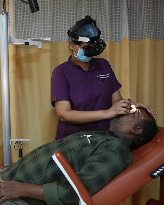
Every patient who visits for a comprehensive eye checkup receives a dilated fundus examination — ensuring retinal disease is detected at the earliest possible stage, when advanced eye treatment options are most effective.
Diabetic Retinopathy — The Leading Cause of Preventable Blindness in Working-Age Adults
Causes no symptoms in its early stages — by the time vision blurs, significant damage may already exist.
Diabetic retinopathy is the most common retinal complication of diabetes. It develops when chronically elevated blood sugar damages the tiny blood vessels that supply the retina, causing them to leak, swell, or grow abnormally. This silent progression is precisely why regular comprehensive eye checkups — including dilated retinal examination — are essential for every diabetic patient, even when vision feels normal.
Non-Proliferative (NPDR)
The early stage, in which retinal blood vessels begin to leak fluid or bleed into retinal tissue. Patients may have no symptoms.
Vision is affected when leakage involves the central macula — diabetic macular edema (DME), the most common cause of vision loss in diabetic patients.
Proliferative (PDR)
The advanced stage, in which the oxygen-starved retina signals the growth of fragile new blood vessels (neovascularisation).
These abnormal vessels bleed easily, causing vitreous haemorrhage and, in severe cases, tractional retinal detachment — requiring prompt treatment.
What Are the Symptoms of Diabetic Retinopathy?
- Blurred or fluctuating vision, particularly after meals
- Dark spots, floaters, or cobweb-like shadows
- Difficulty seeing in low light conditions
- Sudden loss of vision — a sign of vitreous haemorrhage requiring emergency care
- Distorted central vision — straight lines appearing wavy or curved
How Does MediVision Treat Diabetic Retinopathy?
Observation & Risk Factor Control
For very early NPDR, optimising blood sugar, blood pressure, and lipid control alongside 6-monthly retina monitoring.
Intravitreal Anti-VEGF Injections
The primary treatment for diabetic macular edema — reduces swelling by blocking the growth factor driving abnormal vessels.
Laser Photocoagulation
Focal or grid laser reduces macular edema. Pan-retinal photocoagulation treats proliferative disease by reducing ischaemic drive.
Minimally Invasive Vitreoretinal Surgery
For advanced PDR with non-clearing haemorrhage or tractional detachment — performed using the most advanced small-gauge systems available.
Age-Related Macular Degeneration (AMD) — Protecting Your Central Vision
The leading cause of central vision loss in adults over 60.
AMD affects the macula — the central retina responsible for sharp, detailed vision — and can progressively impair reading, face recognition, and all tasks requiring fine visual acuity. AMD exists in two forms, and both require regular retina care monitoring.
Dry AMD (Non-Exudative)
The more common form, accounting for approximately 85–90% of all AMD cases. Gradual accumulation of drusen beneath the retina and progressive atrophy of the macular pigment epithelium.
- Close monitoring with serial OCT imaging
- AREDS2 nutritional supplementation
- Home monitoring with an Amsler grid
Wet AMD (Neovascular)
The less common but more rapidly destructive form. Abnormal new blood vessels grow beneath the macula, leaking fluid and blood that cause sudden, severe central vision loss.
- Urgent intravitreal Anti-VEGF injections
- Regular injection cycles, OCT-guided
- Delays of even weeks materially affect outcomes
What Are the Symptoms of AMD?
- Gradual blurring of central vision
- Straight lines appearing wavy, bent, or distorted
- A grey, dark, or blank area in the centre of vision
- Colours appearing less vivid or washed out
- Difficulty adapting from bright to dim light
Retinal Detachment & Retinal Tears — A Sight-Threatening Emergency Requiring Urgent Retina Care
The retina begins to die within hours of detaching. This is one of the most time-critical emergencies in eye care.
When the retina separates from the underlying retinal pigment epithelium and choroid, it is deprived of its blood supply. Without prompt surgical retina care, permanent blindness in the affected eye is the outcome. Retinal tears typically occur when the vitreous gel shrinks and pulls away from the retinal surface with age — in some patients, this pulls hard enough to tear the retina, allowing fluid to seep beneath it and lift it away.
What Causes Retinal Tears and Detachment?
High Myopia
The most significant risk factor. Highly myopic eyes are longer than average, placing mechanical stress on the peripheral retina.
Previous Eye Surgery
Including cataract treatment and other intraocular procedures.
Eye Trauma or Injury
Direct impact can cause immediate retinal tears or detachment.
Family or Prior History
Family history of retinal detachment, or a previous detachment in the other eye.
Warning Symptoms — Assess the Same Day, Do Not Wait
- Sudden increase in floaters — particularly a shower of new floaters
- Flashes of light in the peripheral visual field
- A curtain, shadow, or dark area across part of the visual field
- Sudden blurring of vision
How Does MediVision Treat Retinal Tears and Detachment?
Barrage Laser Photocoagulation
For tears without detachment. Laser creates a strong adhesion around the tear, preventing fluid passing beneath the retina — a quick, in-clinic procedure.
MIVS — Pars Plana Vitrectomy
For detachment. Our surgeons remove the vitreous, drain subretinal fluid, and reattach the retina using laser and gas or silicone oil tamponade through micro-incisions.
Scleral Buckling
A silicone band sutured around the outside of the eye to indent the scleral wall and support the detached retina from outside, used in selected cases.
Other Retinal Conditions We Diagnose and Treat
From vein blockages to premature-birth screening — the full spectrum of retinal disease under one roof.
Retinal Vein Occlusion (RVO)
RVO occurs when a vein draining blood from the retina becomes blocked, causing blood and fluid to accumulate — most commonly associated with hypertension, diabetes, and elevated cholesterol. Central RVO blocks the main retinal vein, causing widespread haemorrhage affecting central and peripheral vision, and can lead to neovascular glaucoma in severe cases. Branch RVO blocks a smaller vein, typically causing sector visual field loss. Treatment includes intravitreal Anti-VEGF injections, intravitreal steroid implants for refractory cases, pan-retinal laser for proliferative disease, and vitreoretinal surgery for complications.
Central Serous Chorioretinopathy (CSCR)
Fluid accumulates beneath the macula, causing a localised serous detachment that blurs and distorts central vision — most common in young to middle-aged adults, particularly men, and associated with stress and corticosteroid use. Symptoms include blurred central vision, visual distortion, and altered colour perception. Most acute cases resolve within 3–4 months with observation; chronic CSCR (beyond 4 months) may require laser photocoagulation, photodynamic therapy, or oral mineralocorticoid antagonists.
Retinopathy of Prematurity (ROP)
A potentially blinding condition affecting premature and low-birth-weight babies, whose retinal blood vessels have not completed normal development at birth. ROP screening is required for babies with birth weight below 1500g, gestational age of 32 weeks or less, or an unstable clinical course at the neonatologist's discretion. Treatment ranges from close observation for mild disease, to laser photocoagulation for threshold ROP, intravitreal Anti-VEGF injections for aggressive posterior disease, and specialised vitreoretinal surgery for advanced stage 4–5 disease.
Why Does Early Retina Care Save Vision — And Change Outcomes Forever?
Retinal cells cannot regenerate. Early detection is your most powerful tool against vision loss.
The retina is unforgiving. Once retinal tissue dies — from ischaemia, detachment, or progressive disease — the vision it supported cannot be recovered. This biological reality makes early retina care not just beneficial but absolutely essential.
What Does Early Retina Care Achieve?
Prevents Irreversible Vision Loss
A retinal tear treated with barrage laser before detachment requires a 15-minute in-clinic procedure. A full detachment requires surgery, weeks of recovery, and often some permanent loss even with success.
Preserves Central Vision in AMD
Wet AMD causes rapid central vision loss within weeks if untreated. Anti-VEGF therapy promptly stabilises vision in most patients when started without delay.
Protects Working-Age Vision in Diabetes
Diabetic retinopathy is the leading cause of blindness in working-age adults globally. Regular 6-monthly monitoring is the most effective preventive strategy available.
Identifies Systemic Disease
A checkup revealing retinal haemorrhages or vascular changes may be the first indication of uncontrolled diabetes, hypertension, or cardiovascular disease.
Gives ROP Babies a Fair Chance
Undetected and untreated ROP causes irreversible blindness. Timely screening gives every at-risk premature baby the best possible chance of normal visual development.
Recommended Retina Care Schedule
| Patient Group | Recommended Frequency |
|---|---|
| Adults over 40, no risk factors | Annually — dilated fundus exam |
| Diabetic patients | Every 6 months minimum |
| Hypertensive patients | Annually |
| High myopia (above -6.00D) | Every 6–12 months |
| AMD patients (monitored) | Per retina specialist schedule |
| Post-retinal detachment repair | 1 week, 1 month, 3 months, then annually |
| Premature babies (ROP risk) | Per neonatal screening protocol |
Source: American Academy of Ophthalmology retinal examination guidelines.
What Advanced Technology Does MediVision Use for Retina Care?
Precise retina care demands the most advanced diagnostic and surgical tools.
OCT & OCT Angiography (OCTA)
The most important diagnostic technology in modern retina care — micron-resolution cross-sectional imaging of every retinal layer, plus non-invasive blood flow imaging with OCTA.
Fundus Fluorescein Angiography (FFA)
Dye-based retinal vascular imaging — essential for identifying leakage, ischaemia, and neovascularisation in diabetic retinopathy, RVO, and AMD.
Wide-Field Fundus Imaging
Ultra-wide field photography captures up to 200 degrees of the retinal surface in a single image, documenting far-peripheral disease and post-laser treatment zones.
Minimally Invasive Vitreoretinal Systems
Small-gauge (23G, 25G, 27G) MIVS platforms — the most advanced surgical technology for retinal detachment repair and complex diabetic eye treatment.
Laser Delivery Systems
Multi-wavelength platforms delivering precisely calibrated photocoagulation for diabetic retinopathy, RVO, barrage treatment, and CSCR — with pattern laser for faster, more comfortable sessions.
How Does Retina Care Work at MediVision? Your Step-by-Step Journey
From your first call to lifelong monitoring — every step is supported.
Book a Retina Consultation or Eye Checkup
If you have sudden new floaters, flashes, or a curtain across your vision, contact us urgently the same day. For routine monitoring, we schedule your checkup at the earliest convenient slot.
Comprehensive Eye Checkup & Retinal Examination
A full exam — visual acuity, intraocular pressure, anterior segment assessment, and dilated fundus examination — plus OCT, FFA, or fundus photography where clinically indicated.
Diagnosis & Advanced Eye Treatment Plan
Your retina specialist explains the diagnosis, disease stage, and recommended plan in clear language — expected outcomes, timelines, and costs, following NABH consent standards.
Advanced Eye Treatment — Medical or Surgical
From close observation, to same-day intravitreal injections, to laser photocoagulation, to planned vitreoretinal surgery — managed within one integrated system.
Post-Treatment Monitoring & Comprehensive Eye Care
Structured follow-up — OCT monitoring after injections, post-laser review, post-surgical assessment at 1 day, 1 week, 1 month, and 3 months — as part of your ongoing relationship with us.
Why Should You Choose MediVision for Retina Care and Vitreoretinal Surgery?
Complete Range of Retina Care
From diagnostic imaging and intravitreal injections to laser, MIVS, ROP screening, and SFIOL implantation — every retinal condition managed without external referral.
34+ Years, 5.25 Lakh+ Patients
Dedicated subspecialty training across the full range of retinal conditions — from diabetic screening to complex vitreoretinal surgery for advanced disease.
Equipped for Retinal Emergencies
Acute retinal detachment, sudden vitreous haemorrhage, and urgent ROP are managed with priority — patients with detachment symptoms are assessed the same day.
Manages Co-Existing Conditions
Our retina specialists work within a team covering glaucoma, cataract, cornea, and pediatric eye care — for truly integrated comprehensive eye care.
Diabetic Retina Care Programme
Dedicated diabetic eye screening with 6-monthly checkups, OCT and FFA imaging, and injection therapy — one of our highest-impact services.
Your Retinal Health Cannot Wait — Expert Retina Care Is One Call Away.
Retinal disease is silent, progressive, and — in most cases — treatable when caught early. At MediVision Eye Care Centre, our NABH-accredited eye care hospital offers the complete range of retina care services, from routine diabetic eye screening to complex vitreoretinal surgery, across 4 centres, delivered by dedicated retina specialist doctors.
